Taub Institute: Genomics Core
AN NIA-FUNDED ALZHEIMER'S DISEASE RESEARCH CENTER
top
make an appointment

Featured Research

13th ANNUAL TAUB INSTITUTE
RESEARCH RETREAT
November 2023

13th Annual Taub Institute Research Retreat Group Photo

View Archive [close menu]

» 12th ANNUAL TAUB INSTITUTE RESEARCH RETREAT, November 15 2022

» Miguel Arce RenterĂ­a, PhD

» Patrick Lao, PhD

» Caghan Kizil, MSc, PhD

» Kapil V. Ramachandran, PhD

» Radhika Jagannathan, MD, PhD

» Taub Institute Grants: New in 2020

» Spotlight on STAR U: An Interview with Program Directors Drs. Adam Brickman and Stephanie Cosentino, and Program Coordinator Kiana Chan

» ADRC Update: An Interview with Dr. Scott Small

» Chasing Therapeutics: From Neurodegeneration to Sars-CoV-2

» Tal Nuriel, PhD

» Philip L. De Jager, MD, PhD

» Sabrina Simoes, PhD

» 10th ANNUAL TAUB INSTITUTE RESEARCH RETREAT, November 2019

» A Q&A with Brain Donation Coordinator Scott Reid

» Marta Olah, PhD

» Collaboratory on Research Definitions for Reserve and Resilience in Cognitive Aging and Dementia

» Hans-Ulrich Klein, PhD

» Vilas Menon, PhD

» Gunnar Hargus, MD, PhD

» Wassim Elyaman, PhD

» Elizabeth M. Bradshaw, PhD

» Falak Sher, PhD

» Alejandro Chavez, MD, PhD

» Center of Excellence for Alzheimer's Disease (CEAD) at Columbia University Irving Medical Center

» 9th ANNUAL TAUB INSTITUTE RESEARCH RETREAT, November 2018

» Estela Area Gomez, PhD

» Columbia University Alzheimer's Disease Research Center (ADRC) 2018-19 Pilot Grant Awardees

» Luke E. Berchowitz, PhD

» Catherine Marquer, PhD

» Inbal Israely, PhD

» Laura Beth McIntire, PhD

» Abid Hussaini, PhD

» Yian Gu, MD, MS, PhD

» 8th ANNUAL TAUB INSTITUTE RESEARCH RETREAT, November 2017

» Frank Provenzano, PhD

» James M. Noble, MD, MS

» Ronald K. H. Liem, PhD

» Natura Myeku, PhD

» Christiane Reitz, MD, PhD

» Qolamreza R. Razlighi, PhD: Quantitative Neuroimaging Laboratory

» Nicole Schupf, PhD

» Sandra Barral Rodriguez, PhD

» Wai Haung Yu, PhD

» Giuseppe Tosto, MD, PhD

» Catherine L. Clelland, MS, PhD

» 7th ANNUAL TAUB INSTITUTE RESEARCH RETREAT, October 2016

» Andrew A. Sproul, PhD

» William C. Kreisl, MD

» Badri N. Vardarajan, PhD, MS

» Diego E. Berman, PhD

» Joseph H. Lee, DrPH

» Dr. Yaakov Stern: the Concept of Cognitive Reserve

» The Alzheimer's Disease Research Center at Columbia University Celebrates 25 Years

» Stephanie Cosentino, PhD

» Edward D. Huey, MD

» Jennifer J. Manly, PhD

» Lawrence S. Honig, MD, PhD, FAAN

» 6th ANNUAL TAUB INSTITUTE RESEARCH RETREAT, October 2015

» Andrew Teich, MD, PhD

» Ismael Santa-Maria Perez, PhD

» Christian Habeck, PhD

» Roger Lefort, PhD

» Clarissa Waites, PhD

» Francesca Bartolini, PhD

» Tae-Wan Kim, PhD

» Carol M. Troy, MD, PhD

» Adam M. Brickman, PhD

» Gil Di Paolo, PhD

» 5th ANNUAL TAUB INSTITUTE RESEARCH RETREAT, October 2014

» Asa Abeliovich, MD, PhD

» Lorraine N. Clark, PhD

» Ulrich Hengst, PhD

» Yaakov Stern, PhD: Cognitive Neuroscience of Aging Laboratory

» Ottavio Arancio, MD, PhD

» Karen S. Marder, MD, MPH

» Scott Small Laboratory

» Michael Shelanski Laboratory

» Richard Mayeux, MD, MSc: Laboratory for Genetic Epidemiology
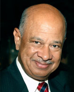
» Karen Duff Laboratory


Aβ and Tau Oligomers Cause Profound Changes in Transmitter Release Via Presynaptic APP

Ottavio Arancio, MD, PhD
Professor of Pathology and Cell Biology and of Medicine (in the Taub Institute)

Amyloid precursor protein (APP) is a transmembrane protein expressed at the synapse throughout life. In absence of APP, extracellular AĂź- and tau-oligomers no longer impair memory and its synaptic surrogate, long-term potentiation (LTP). Synapses include pre- and post-synaptic compartments. However, the relative role of pre- vs. post-synaptic APP at the CA3-CA1 hippocampal synapse in the AĂź- and tau-oligomer-induced damage of memory and LTP is not known.


Deep Learning on Clinical Neuroimaging and Neuropathology in Lewy Body Dementia and Alzheimer’s Disease

Frank Provenzano, PhD
Assistant Professor of Neurological Sciences (in Neurology and in the Taub Institute)

Neuroimaging signatures of Alzheimer’s Disease and related dementia’s have been well-explored in research settings largely using fixed and harmonized protocols. Two limitations of these datasets is the availability of neuropathology to define and quantify disease burden and confirm clinical diagnosis as well as the lack of translation to clinical datasets. Building off existing research identifying longitudinal neuroimaging signatures, we demonstrate a strategy to explore how we can use machine learning methods to clarify and classify ante-mortem MRI. We show the process where we use a collection of both research and clinical MRI machine learning approaches to accurately Alzheimer’s Disease with and without Lewy Body Dementia more accurately than clinical diagnosis.


Translational Genomics Approaches for Functional Investigation of Alzheimer’s Related Genes in Zebrafish

Caghan Kizil, PhD, MSc
Associate Professor of Neurological Sciences (in Neurology and in the Taub Institute)

Zebrafish stands as a promising model for Alzheimer's disease (AD) research with its biological validity, disease modeling capabilities, translational genomics approaches, suitability for therapeutic screening, and ethical advantages. Zebrafish shares high gene and protein similarity with humans, making it an ideal platform for neurodevelopmental, neurological, and behavioral investigations. Leveraging developed genetic tools and cost-effective maintenance, we have established "zebrafish-to-human" and "human-to-zebrafish" approaches for translational genomics, including the use of CRISPR/Cas9 to investigate AD-related gene function. In one instance, we utilized CRISPR/Cas9 to create a zebrafish knockout model for abca7, an AD-risk gene homologous to human ABCA7. This model demonstrated reduced astroglial proliferation, synaptic density, and microglial abundance in response to Aβ42, uncovering an abca7-dependent neuro-glial crosstalk mediated by neuropeptide Y (NPY) signaling. Clinical data and genetic variants in NPY, BDNF, and NGFR further highlighted the role of ABCA7-dependent NPY signaling in synaptic integrity and brain resilience. Additionally, we explored the protective mechanisms against APOEε4, a major AD risk factor in human cohorts and zebrafish. We identified protective variants exclusive to APOEε4 carriers, which were associated with extracellular matrix-related processes. In zebrafish, loss-of-function mutations in one ECM component ameliorated AD related cellular changes such as gliosis, gliovascular remodeling, and microglial response. I will discuss these two exemplary cases in my presentation.


Characterizing Glial Signatures in the Human Neuritic Plaque Microenvironment Using Spatial Transcriptomics

Mariko Taga, PhD
Assistant Professor of Neurological Sciences (in Neurology and in the Taub Institute)

Neuroinflammation is one of the consistent features of Alzheimer’s Disease (AD). Recent technological advances in single-nucleus RNA-sequencing (snRNA-seq) revealed a large diversity of molecularly-defined, immunocompetent human glial cell populations and identified specific glial cell subpopulations associated with AD pathologies. However, due to the loss of spatial information, identifying glial signatures spatially associated with AD pathologies is challenging.

In our study, Spatial Transcriptomic (ST) combined with Immunofluorescence (IF) was conducted on 32 sections from the dorsolateral prefrontal cortex of 15 AD and 2 control subjects. We compared the transcriptional profile of regions within 150ÎĽm of neuritic plaques to those located more than 500ÎĽm away from the plaques. This approach allowed us to identify several glial genes that were either upregulated or downregulated within the neuritic plaque microenvironment, including SERPINA3 and metallothionein genes.

To further investigate the state of glial cells within this neuritic plaque microenvironment, we employed Cell2location to deconvolute the spatial transcriptomics data. We used single-nucleus RNA sequencing data generated from 450 samples from ROS/MAP subjects to construct a single nucleus reference of transcription profiles with cell state annotations. Our analysis revealed that a specific astrocyte cluster expressing SERPINA3 is enriched in the neuritic plaque microenvironment as well as a DAM-like microglia cluster. Modulating the expression of these specific genes, which characterize glial signatures spatially associated with neuritic plaques, may provide insights into the function of glial cells in plaque clearance and the progression of pathology.


New Developments in Genetics of AD in Diverse Populations

Christiane Reitz, MD, PhD
Associate Professor of Neurology and Epidemiology (in the Gertrude H. Sergievsky Center and the Taub Institute)

Limited ancestral diversity has impaired our ability to detect risk variants more prevalent in non-European ancestry groups in genome-wide association studies (GWAS). As part of the Alzheimer’s Disease Genetics Consortium we constructed and analyzed multi-ancestry and African ancestry GWAS datasets to identify novel shared and ancestry-specific AD susceptibility loci. The multi-ancestry GWAS in 37,382 non-Hispanic White (NHW), 6,728 African American, 8,899 Hispanic (HIS), and 3,232 East Asian individuals identified 13 loci with cross-ancestry associations including known loci at/near CR1, BIN1, TREM2, CD2AP, PTK2B, CLU, SHARPIN, MS4A6A, PICALM, ABCA7, APOE and two novel loci not previously reported at 11p12 (LRRC4C) and 12q24.13 (LHX5-AS1), in addition to three ancestry-specific loci near PTPRK and GRB14 in Hispanics, and KIAA0825 in non-Hispanic Whites. Genes at these novel loci have known roles in neuronal development (LRRC4C, LHX5-AS1, and PTPRK) and insulin receptor activity regulation (GRB14). The independent African ancestry GWAS based on the African Genome Resources reference panel identified a novel AD risk locus in MPDZ involved in synaptic transmission. These findings underline the importance of using traditionally underrepresented populations for gene discovery, even with smaller sample sizes.


Cognitive Effects of Bilingualism: A Global Perspective

Miguel Arce RenterĂ­a, PhD
Assistant Professor of Neuropsychology (in Neurology)

Bilingualism is associated with a “cognitive advantage” such that bilinguals demonstrate better cognitive test performance compared to monolinguals. However, most of the literature on a bilingual cognitive advantage has largely relied on study populations from high income countries, whereas the vast majority of bilingual/multilingual adults reside in low-and-middle income countries (LMICs). In addition, prior studies on the cognitive effects of bilingualism treat bilingual adults as a monolithic group when in fact bilinguals demonstrate great within-group heterogeneity in both linguistic and sociocultural backgrounds. For instance, bilingual adults can differ on their age of acquisition, level or proficiency, frequency of use, types and number of languages known, as well as the sociocultural context in which these languages are used. To improve our understanding of the role of bilingualism and cognition, research that deconstructs bilingualism and its sociocultural factors across both high and LMICs is needed. To highlight this approach, we will show three different ongoing studies evaluating bilingualism and cognition across: 1) Spanish-English bilingual Latinos residing in the US, 2) Spanish-English and Spanish-Indigenous Language bilingual adults residing in Mexico, and 3) multilingual adults residing in India.


Neuroimaging of Vascular and Inflammatory Pathways in Alzheimer’s Disease

Patrick Lao, PhD
Assistant Professor of Neurological Sciences (in Neurology and in the Gertrude H. Sergievsky Center)

Amyloid and tau PET imaging with structural MRI can stage Alzheimer’s disease (AD), but the pathways by which tau aggregates, spreads, and leads to downstream neurodegeneration remain unclear. Cerebrovascular disease, particularly small vessel disease, may emerge as a part of the disease process rather than being a simple comorbidity, and may be locally associated with phosphorylated tau, independently of amyloid. Neuroinflammation, particularly microglia, may mediate tau spreading across key AD brain regions and downstream neurodegenerative and cognitive consequences. Together, vascular and inflammatory pathways provide a more complete understanding of the amyloid-tau-neurodegeneration framework.


Understanding the Role of the Ubiquitin Proteasome System in Alzheimer’s Disease: Pathways and Potential Therapies

Natura Myeku, PhD
Assistant Professor of Pathology and Cell Biology (in the Taub Institute) at the CUMC

Neurodegenerative diseases (NDs), particularly Alzheimer's disease (AD), are characterized by the accumulation of disease-specific proteins in the brain, leading to cellular dysfunction, synapse loss, and degeneration. A major constituent of these aggregates is ubiquitin, suggesting that disrupted ubiquitin-proteasome-mediated degradation could be a driving force behind NDs or a consequence of their pathogenesis. While the proteasome's role in regulating the ubiquitin-proteasome system (UPS) in AD and other NDs has historically been underestimated, recent studies acknowledge its crucial role in protein homeostasis. Our research aims to understand the mechanistic cascade of protein breakdown failure by the 26S proteasome in AD using integrated multi-omics and functional approaches. Though the regular 26S proteasome is found in all cells, a different form, the inducible proteasome, is located exclusively in the brain's glial cells. Its exact role in AD is still uncertain. It's undetermined whether these inducible immunoproteasomes are protective or if their presence accelerates AD's development. Considering the functional diversity of immunoproteasomes, we think that it is crucial to elucidate the true nature of immunoproteasomes in AD since their biogenesis and activity can be exploited further by pharmacological means for therapeutic purposes. Using novel transgenic crosses and current transcriptomic and proteomic technology, we aim to investigate whether immunoproteasome deficiency can exacerbate tau and amyloid pathology.


Association Between APOE-ε4 Allele and the Risk of Alzheimer’s Disease Across Populations with Diverse Genetic Ancestries

Sandra Barral Rodriguez, PhD
Associate Professor of Neurogenetics (in Neurology, the Gertrude H. Sergievsky Center and the Taub Institute) at the CUMC

Alzheimer’s disease and Related Dementias (ADRD) represents the most common form of dementia worldwide. APOE-ε4 allele continues to be the strongest and most replicated genetic risk factor reported in the non-Hispanic White population. A small number of studies have addressed the relationship between APOE and ADRD risk in non-White populations, and the results have not followed a consistent pattern so far. Extending genetic studies to include large samples of diverse and admixed populations is critical for gaining insight into the variation of genetic risk among populations with diverse ancestries.

Our study included a total sample of 8,476 individuals from six studies and four different populations: Mexicans, Mexican-Americans, Peruvians, and Caribbean-Hispanics. We showed a heterogenous genetic ancestral composition across the cohorts, with the Peruvian population exhibiting the highest Native American ancestry proportion followed by Mexicans and lastly Caribbean Hispanics. The strongest association between APOE-ε4 and ADRD risk was observed in Peruvians, while ε4-allele effect proving similar for the other cohorts. Meta-analyses also found a nominally significant protective effect against ADRD risk for the ε2 allele.

Our results showed a heterogenous risk for ADRD from the APOE isoforms across Hispanics. To our knowledge, this is the largest collection of Hispanic individuals with different ancestry background phenotyped for ADRD.


Investigating the Role of APOE4 in Alzheimer's Disease Pathogenesis

Tal Nuriel, PhD
Assistant Professor of Pathology and Cell Biology (in the Taub Institute) at the CUMC

Carriers of the apolipoprotein E (APOE) ε4 gene are at a significantly increased risk for developing Alzheimer’s disease (AD). Although numerous theories have been proposed, the cause of this association remains unclear. Our research has uncovered novel effects of APOE4 expression on important processes in the brain, including the endosomal-lysosomal system, bioenergetic regulation, and neuronal activity. However, substantial questions remain about when, where and how these systems are affected by differential APOE isoform expression, as well as the impact of these pathway-level effects on AD pathology and also the human relevance of these effects. In order to answer these questions and gain a more comprehensive understanding of how differential APOE isoform expression affects vital brain processes and pathways, our lab is conducting numerous innovative research studies utilizing mouse models, human tissues and data, and cell lines. In addition, we utilize cutting-edge techniques, such as spatial omics, MRI, and in vivo live imaging to facilitate these investigations. In this presentation, I will update my Taub colleagues on the recent progress made in these studies, and what these results tell us about how APOE4 expression may accelerate AD pathogenesis. We anticipate that the completed results from these studies will greatly increase our understanding of how APOE4 affects an individual’s susceptibility to AD, potentially leading to new preventative and therapeutic strategies for AD, especially among APOE4 carriers.


KEYNOTE LECTURE


Genetic Risk for Alzheimer’s Disease: What this Tells us About Mechanism of Disease

Alison M. Goate, DPhil
Jean C. & James W. Crystal Professor and Chair
Director, Ronald M. Loeb Center for Alzheimer's disease
Dept. of Genetics & Genomic Sciences, Icahn Genomics Institute
Icahn School of Medicine at Mount Sinai

Alzheimer’s disease is the most common form of dementia affecting more than 6.7 million Americans age 65 years and older. It is characterized clinically by slowly progressive memory loss and neuropathologically by the presence of extracellular ß-amyloid (Aß) plaques, intracellular accumulation of neurofibrillary tangles and the presence of neurodegeneration and gliosis. Studies in genetic and sporadic forms of AD have demonstrated that both forms of the disease are characterized by a prodromal phase, lasting up to two decades, in which plaques and tangles accumulate leading to neurodegeneration. Inherited forms of AD are associated with mutations in the amyloid precursor protein (APP) gene or the presenilins (1 & 2) and directly implicate APP metabolism in disease pathogenesis. During the last year several antibody-based therapies targeting ß-amyloid have been approved by the FDA. However, although Aß accumulates in sporadic forms of the disease other mechanisms are likely to be important. The most common genetic risk factor in late onset AD, is apolipoprotein E (APOE) genotype. Both risk and protective alleles have been identified enabling mechanistic studies of the role of APOE in AD risk. Genome-wide association studies have implicated close to one hundred other loci in AD risk and integrative genomic studies demonstrate that these common alleles are largely non-coding and reside in microglial enhancers. Genomic, epigenomic and functional studies demonstrate that these risk genes influence microglial state and their response to Aß and/or neurodegeneration. Rare coding variants in TREM2, PLCG2 and ABI3 also highlight the importance of microglia and implicated TREM2 signaling as a central biochemical response of microglia to Aß or damage caused by Aß in the AD brain.


bottom bar